
MOM®
Movable Objective Microscope®
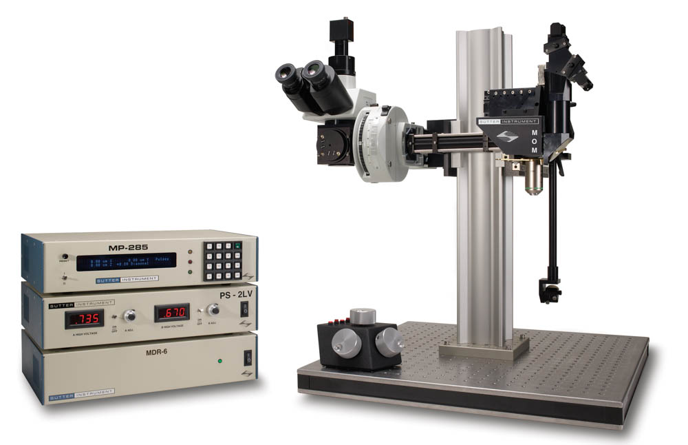
The Movable Objective Microscope® (MOM®) is a two- or thee-photon microscope capable of imaging deep within living specimens when combined with an appropriate laser. The Sutter MOM was the first scope to provide 3-dimensional objective movement and rotation allowing the specimen to remain horizontal and stationary. Many highly regarded imaging laboratories around the world use the Sutter MOM and we constantly work with our customers to adapt the design for their changing needs.
Watch the video describing MOM imaging and photostimulation beam paths
MOM Opto-mechanical Design
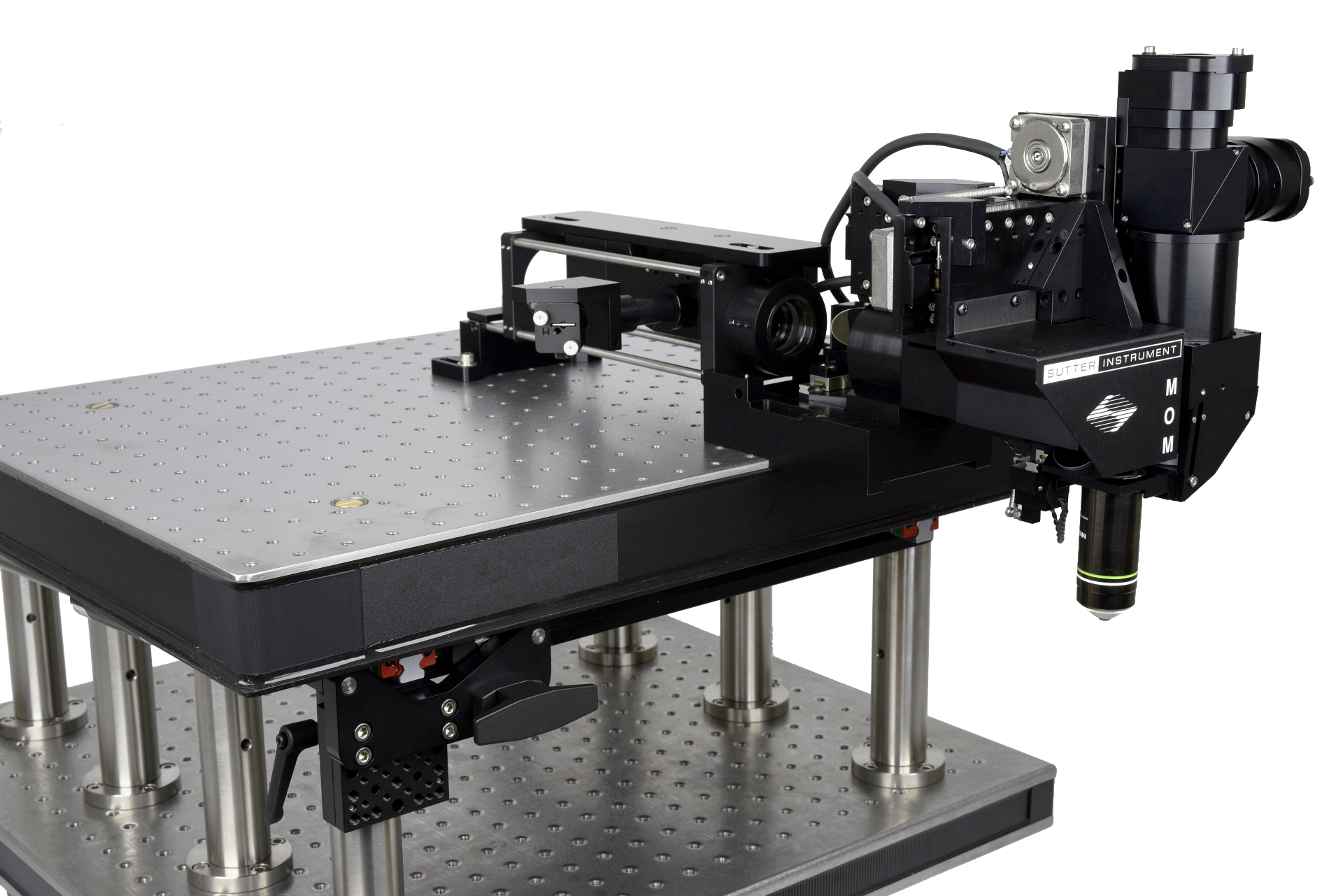
The MOM consists of two independent microscopes. The wide-field half of the microscope consists of an Olympus vertical illuminator, Sutter Xenon arc lamp and camera mount to provide standard epifluorescence. The two-photon side of the microscope provides the optical pathway for guiding the excitation laser light from the table up into the scanning galvanometric mirrors and then expanding the beam through the scan lens and directing into the back of the objective. Following twophoton excitation, the emitted photons are directed by a dichroic mirror immediately above the objective into the detection pathway. The main body of the microscope moves backwards on a rail system allowing easy access to the specimen prior to imaging.
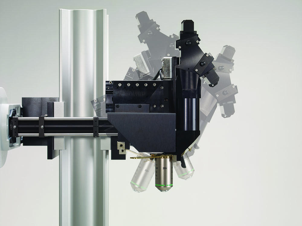
The objective translates in X, Y and Z as well as rotates around the X axis. Two moving mirrors allow the microscope to maintain efficient delivery of the excitation light to the back aperture of the objective regardless of movement or orientation. The X, Y and Z movements used are the same as that in our MP-285 micromanipulator so you know the movements are smooth, fine in scale, drift-free and highly reproducible. These movements permit Z-stacks and mosaic images of large regions of tissue to be recorded without the need for a moving stage.
The horizontal light path allows for rotation of the objective away from the standard vertical position. As a result of this rotation, the MOM can easily be converted from an upright to an inverted microscope and the objective positioned from 0 to 180 degrees. This positional freedom permits the imaging of non-horizontal surfaces and volumes.
MOM Scanning Systems
During the last 10 years, scanning systems for multiphoton microscopes have changed in several ways. Large aperture, high NA objectives became available and thus required larger aperture scanners. Resonant scanner technology allowed faster imaging. Two-photon scopes now include both resonant-galvo and resonant galvo-galvo systems. The Sutter MOM developed in parallel with these changes and new technology can be bolted into older, existing scopes with minimal changes. Many original scopes with 3 mm galvo scanners have been upgraded to either 6 mm galvo scanners or resonant/galvo scanners. As an example, the Vidrio RMR scanner (a resonant galvo-galvo scanner system) can be purchased as part of any new MOM system or retrofit into existing MOM scopes.
Imaging Software
Starting in 2011, Sutter began offering the MOM Computer System and Software (MCS). Before this software package was developed, most users relied on ScanImage or MPScope to generate scanned images. Customers valued the fact that the MOM would operate with open source freewares, however, there seemed to also be a market for a commercial package. MCS continues to offer a simple, easy to use package available at a price that compares with other commercial and freeware packages. MScan 3.0, the latest version, is Windows 10 compatible. A recent publication takes advantage of the long (1-2 hour) data files that can be captured in the MCS proprietary data file structure. (reference Kuhn, 2020).
The MOM® has always been compatible with ScanImage freeware, the two-photon imaging software developed by Karel Svoboda and collaborators. One of the reasons the MOM platform exists in its present form is the strong support from the ScanImage community. In 2014, Vidrio became the principle vehicle for support and new development of ScanImage. Sutter is happy to make Vidrio ScanImage Premium available to customers who wish premium support and the latest features. ScanImage Basic is available as an entry level system with a year of support included. ScanImage freeware is still available but does not include support. Sutter provides packages that include the necessary data acquisition hardware to couple the MOM and other scanning microscopes to ScanImage Premium, ScanImage Basic or the freeware version. We also sell Vidrio's hardware line including ScanImage ready computers, the vDAQ acquisiton system and the RMR scanner.
Sutter MOM packages include all of the equipment (less the laser and objective) needed for a complete imaging system:
- Scan lens and tube lens appropriate for two- or three-photon imaging
- Cambridge Technology XY galvonometric (3 mm or 6 mm) or resonant scanners (resonant-galvo or resonant galvo-galvo systems both with 5 mm mirrors)
- Hamamatsu photomultiplier tubes (PMTs): R6357 multialkali or H10770PA-40 (GaAsP) products. (Other PMTs are available, Sutter is an authorized reseller for Hamamatsu)
- Power supplies for PMTs: Sutter PS-2 (dual channel high-voltage power supply for conventional PMTs) or Sutter PS-2/LV (dual channel low-voltage power supply for H10770PA-40 or other PMTs with built in high voltage). Power supplies can be ordered with remote turn on/shut off for PMT gating
- Hamamatsu, Sigmann, or FEMTO pre-amplifiers
- Data acquisition: National Instruments PXI FPGA, Vidrio vDAQ, or National Instruments PC based Multifunction I/O
- Conoptics Pockels Cells for laser intensity control
- APPLICATIONS
- In vivo two-photon imaging
- In vivo three-photon imaging
- Electrophysiological recording and imaging (culture, large in vivo preparations, etc.)
- Non-horizontal surface microscopy
- Simultaneous retinal stimulation and two-photon microscopy1
- Whole animal imaging
- Immunology
- Embryology
- FEATURES
- Objective moves 22mm in X, Y, and Z
- Objective rotates about optical axis for imaging of non-horizontal surfaces and volumes
- Customizable open platform design
- Cambridge Technology conventional or resonant XY scanners
- Two or four channel detector system with Hamamatsu PMTs and preamplifiers
- Sutter PS-2/ PS-2LV dual channel PMT power supply
- Two- or three-photon compatible scan lens and tube lens
- *New* Light Block keeps visual stimuli, photostimuli, and ambient light out of detector path
- National Instruments and Vidrio Technologies data acquisition systems
1 "Eyecup scope-optical recordings of light stimulus-evoked flourescence signals in the retina", Euler et al, Pflugers Arch, 2008
Sutter Instrument Relayed RGG Scanbox
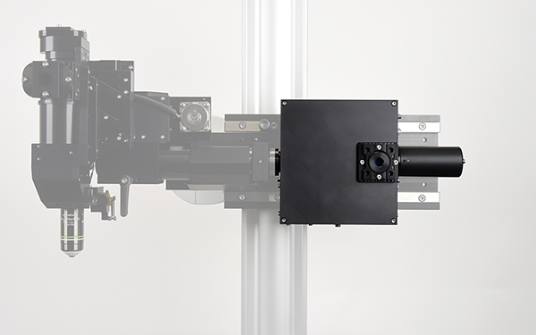
Many neuro-imaging labs are looking for ways to increase the functionality of their standard galvo-galvo (GG) or resonant-galvo (RG) scanning 2-photon microscopes. Upgrading to resonant-galvo-galvo scanning (or RGG scanning) is one way to add new capabilities to your system. The new Sutter RGG scanbox adds new functionality in a compact form factor.
What are the advantages of an RGG scanbox?
1) On a single laser path scope, RGG can function as either a GG or RG pathway. One can do fast scanning to monitor changes in imaged calcium levels using the RG mode (with the second galvo scanner commanded to stay at the neutral position) or it can do slower, high-resolution scanning in GG mode (with the resonant scanner commanded with zero volts to stop resonating).
2) Many experimenters wish to be able to photo-stimulate a portion of the field of view (FOV) or a single part of a cell to photoactivate one or more small areas in sequence followed by fast RG scanning to see how cells react to the photo-stimulation. This can often only be done with two scan paths, one RG and one GG. An RGG scan box allows that to be done with a single scan path keeping the design simpler.
3) Finally, a methodology sometimes referred to as “postage stamp” or “region of interest” (ROI) scanning can be used to limit the areas exposed to excitation light and in certain cases, also allow for faster scanning.
Why would you want to have a Sutter Relayed RGG Scanbox?
1) Like other designs, our RGG beam path relays the resonant scanner center of rotation into the center of rotation of the first galvo. Unlike other designs, we do not rely on a lens-based relay system, so we maintain more power and induce less pulse dispersion than lens based relayed scanners.
2) It was designed to fit the Sutter MOM and thus will also fit the Sutter DF-Scope. The RGG Scanbox will be available in the “Simple 2P” and the BOB-2P microscopes. The relay system keeps the design small (6 X 6 X 2 inches) so it will fit into any number of conventional scanning scopes. We expect a generic Relayed RGG Scanbox to be available soon.
3) You can expect Sutter quality, technical support as well as presale support just like all our other products.
Forget "multiphoton ready"!
The new Sutter 3P-MOM is ready to go
for deep-tissue, three-photon imaging.
Despite the intense interest in three-photon microscopy over the last two years, this is still a relatively nascent field. Neurobiologists doing two photon imaging and microscope manufacturers building two photon microscopes have been eager to make the jump into the technology that will allow experimenters to image deeper into the brain, probably close to twice the sub millimeter depth of the best two photon recordings. We all hoped we could just dial the laser up to 1300 nm and start imaging deeper. After all, Chris Xu made it look so easy!
Three-photon microscopy is at about the same stage as two-photon microscopy was in the late ‘90s. There were a small number of labs doing the necessary work to establish the field. Most made their own microscopes and commercialized systems were not well suited to doing functional imaging in vivo.
Today, with respect to three-photon microscopy, there are a handful of labs that are establishing the field, largely using either homemade microscopes or adapting existing two photon scopes. As yet, there does not seem to be a huge market for de novo commercial platforms, possibly because the technique is more challenging than two photon and possibly because three photon requires a completely different excitation source than two photon with more power concentrated in narrower, taller pulses. In this developing field, Sutter’s Microscope division is exactly where you would expect and want us to be - working with a significant portion of the labs developing three-photon microscopy. Most started with our two photon MOM® design and have converted them to three photon platforms either on their own or with our assistance. Now, Sutter can incorporate those changes into any existing Sutter 2P-MOM. The most important change is to convert the scan and tube lenses to better transmit IR beyond 1700 nm. We have also collaborated to develop a more efficient version of one of our detector paths to do a better job of collecting emission from even deeper focal depths.
In the last two years we have quoted and built a number of three photon MOMs as well as a few that were designed to be “three-photon ready”. We have collaborated with several labs to convert existing 2P-MOMs into 3P platforms.
We are excited to be fully involved in this developing field. Let us help you see what the challenges and benefits of three photon imaging can be. Whether you are just entering the field of multiphoton imaging or are an experienced two photon imager who wishes access to deeper and lower-noise images that three-photon excitation can bring, please contact us.
TECHNICAL SPECIFICATIONS
Travel
22 mm on all three axes
Resolution
MP-285 controller
Low: 0.2 µm/step
High: 0.04 µm/step
MPC-200 controller
0.0625 µm/step
Maximum Speed
MP-285 controller
2.9 mm/sec
MPC-200 controller
5.0 mm/sec
Long Term Stability
1-2 µ/hour
Drive Mechanism
Precision worm gear capstan drive
Communication
MP-285: RS-232 Serial
MPC-200: USB
Electrical
115/230 volts
50/60 Hertz power line
LAMBDA LS 300W XENON ARC LAMP
Lamp Life
1,000 hours (500 hour warranty)
Longer life depends on application
Electrical
115/230 volts
50/60 Hertz power line
PS-2/PS-2LV PMT POWER SUPPLY
Electrical
115/230 volts
MDR-3/MDR-6/MDR-R SCAN DRIVE CONTROLLER
Electrical
115/230 volts
50/60 Hertz power line

![]()
US PRICES > MOM®
International prices vary by country. Please contact your local distributor or Sutter Instrument for a quotation. Prices subject to change without notice.
MOM - BASIC SYSTEM FOR 2-PHOTON MICROSCOPY
Includes Moving Objective Microscope, 2 channel detector with PMTs, preamps and PS-2 power supply, XY scanners with drive electronics, wide field fluorescence unit including vertical illuminator, LB-LS 300
Watt Xenon Arc Lamp, LLG and light guide adapter, C-mount for wide field camera, data acquisition system. Please contact SUTTER for price quotes
| Catalog Number | Description | Price |
| MOM-5MM1 | MOM System with 5mm XY scanners | $ CALL |
|---|---|---|
| MOM-RES-MCS1 | MOM System with Resonant scanners and MCS 3.0 | $ 172,438 |
| MOM-RES-SIP1 | MOM System with Resonant scanners and ScanImage Premium | $ 185,214 |
| MOM-RMR-SIP1 | MOM System with Vidrio RMR scanners, GaAsP PMTs, vDAQ acquisition system and ScanImage Premium | $ 215,762 |
| MOM-3P1 | MOM System with 3P scan and tube lens, 3P detector path, 6 mm galvo scanners, and ScanImage Premium | $ CALL |
US PRICES > ACCESSORIES
| Catalog Number | Description | Price |
| MOM-SETUPKIT-M | Basic table optics for laser routing | $ 2,904 |
|---|---|---|
| MOM-ALIGNTOOL2 | MOM alignment tool | $ 2,487 |
| MOM-LB-WIDE | Light blocking cover excludes ambient and other light from detector path | $ 3,614 |
| MOM-3P-CONVERSION | 3P compatible scan lens and tube lens. Price depends on current MOM configuration | $ CALL |
| MOM-3P-SHORTPATH | Call Sutter for details | $ CALL |
1 Final pricing depends on detector path selected and does not include several devices necessary for a complete 2-photon microscope (i.e. Ti:Sapphire laser, objective, camera, trinocular head, table mount optics).
Please phone Sutter for details.
2 Useful tool for aligning the laser in MOM scopes, especially those with resonant scanners
RELATED PRODUCTS
MCS Data Acquisition System
PRODUCT INFORMATION
Download MOM® Sales Flyer
Download 3P-MOM® Sales Flyer
Download MOM with Neurotar Mobile HomeCage™ Sales Flyer
VIDEOS
WEBINAR: Open Platform Two-Photon Microscopy
MOM scope translation to allow easy access to preparation
Rotation of MOM head to image non-horizontal surfaces
MOM Objective movement in X, Y and Z
Beam Alignment Tutorial for the Movable Objective Microscope
Beam Paths of the Movable Objective Microscope (MOM)
RELATED ARTICLES
Imaging Brains of Mice On The Go Nature Research Custom Media
Clancy K. B., Koralek A. C., Costa R. M., Feldman D. E. & Carmena J.M. (2014). Volitional modulation of optically recorded
calcium signals during neuroprosthetic learning. Nature Neuroscience
Victoria Phoumthipphavong, Florent Barthas, Samantha Hassett, Alex C. Kwan. Longitudinal Effects of Ketamine on Dendritic Architecture In Vivo in the Mouse Medial Frontal Cortex. eNeuro.org DOI: 10.1523/ENEURO.0133-15.2016